Transient bone marrow oedema of the hip also referred as transient osteoporosis of the hip is self limited conditions that improves spontaneously over several months.
Transient osteoporosis of the hip mri.
Mri with t1 and t2 weighted sequences in coronal transverse and sagittal sections was performed in 12 patients with retrospectively confirmed to both at the onset of the disease and later as follow up procedure.
Three patients with transient osteoporosis of the hip underwent magnetic resonance mr imaging.
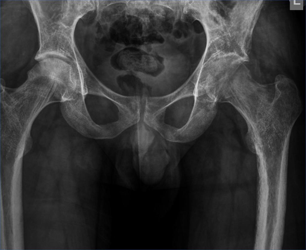
Transient osteoporosis of the hip also known as transient bone marrow edema syndrome of the hip is a self limiting clinical entity of unknown cause although almost certainly a vascular basis and possible overactivity of the sympathetic system exists there is some controversy as to whether transient osteoporosis of the hip represents a very early reversible stage of avascular necrosis.
Toh was first described in 1959 in two women in their 3rd trimesters of pregnancy but now is more commonly seen in middle aged men.
Because of this mri is one of the most useful studies to help diagnose the condition.
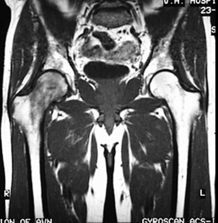
By contrast mri changes in avn of the femoral head is restricted to a smaller.
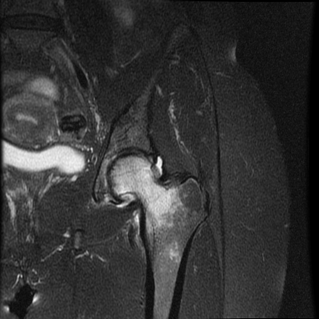
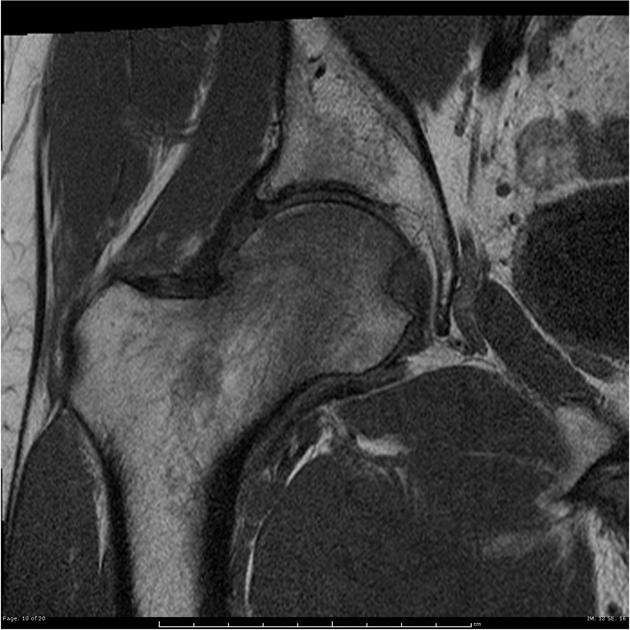
Mri changes in transient osteoporosis of the hip involve the whole femoral head and sometimes extend to the femoral neck and even the trochanteric region with blurred and vague margins.
An mri scan of a hip affected by transient osteoporosis will usually reveal bone marrow edema.
This mri image shows edema surrounding the affected hip.


